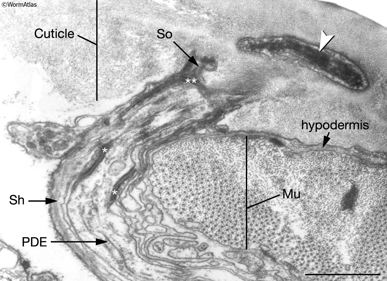
42 transmission electron micrograph labeled
Solved Label the transmission electron micrograph of the - Chegg Label the transmission electron micrograph of the mitochondrion. Matrix granule Mitochondrion Outer membrane Cristae Inner membrane Matrix Reset Zoom Question: Label the transmission electron micrograph of the mitochondrion. Matrix granule Mitochondrion Outer membrane Cristae Inner membrane Matrix Reset Zoom This problem has been solved! (A and B) Electron micrograph of a cell labeled for/5-tubulin followed... Transmission electron microscopy has proven to be a powerful tool in providing information about nanoparticle uptake, biodistribution and relationships with ...
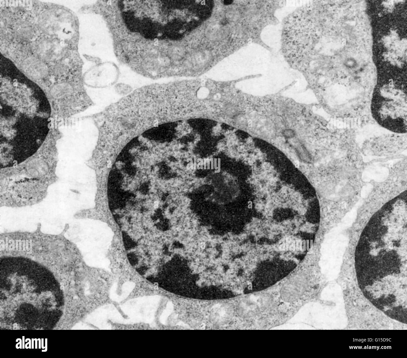
Solved Label the transmission electron micrograph of the - Chegg Transcribed image text: Label the transmission electron micrograph of the cell. 0 Nucleus rences Mitochondrion Heterochromatin Peroxisome Vesicle ULAR bumit Click and drag each label into the correct category to indicate whether it pertains to the cytoplasm or the plasma membrane.
Transmission electron micrograph labeled
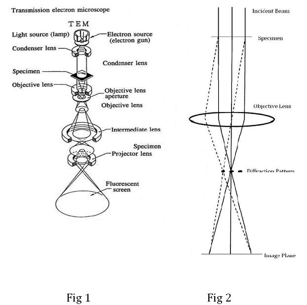
8.2: Transmission Electron Microscopy - Chemistry LibreTexts Transmission electron microscopy (TEM) is a form of microscopy which in which a beam of electrons transmits through an extremely thin specimen, and then interacts with the specimen when passing through it. The formation of images in a TEM can be explained by an optical electron beam diagram in Figure 8.2. 1. Transmission Electron Microscopy as a Powerful Tool to Investigate ... Despite the high resolution (3–20 nm), imaging is restricted to the surface of the sample. ... Transmission electron microscopy (TEM) provides images obtained by ... Labeling the Cell Flashcards | Quizlet Label the transmission electron micrograph of the nucleus. membrane bound organelles golgi apparatus, mitochondrion, lysosome, peroxisome, rough endoplasmic reticulum nonmembrane bound organelles ribosomes, centrosome, proteasomes cytoskeleton includes microfilaments, intermediate filaments, microtubules Identify the highlighted structures
Transmission electron micrograph labeled. Transmission Electron Microscopy | Central Microscopy Research Facility The transmission electron microscope (TEM) operates on many of the same optical principles as the light microscope. The TEM has the added advantage of greater resolution. This increased resolution allows us to study ultrastucture of organelles, viruses and macromolecules. Specially prepared materials samples may also be viewed in the TEM. en.wikipedia.org › wiki › Transmission_electronTransmission electron microscopy DNA sequencing - Wikipedia Transmission electron microscopy (TEM) produces high magnification, high resolution images by passing a beam of electrons through a very thin sample. Whereas atomic resolution has been demonstrated with conventional TEM, further improvement in spatial resolution requires correcting the spherical and chromatic aberrations of the microscope lenses. Label This Transmission Electron Micrograph - Kaiden Brown Label this transmission electron micrograph of relaxed sarcomeres by clicking and dragging the labels to the correct location . Transmission electron microscopy (tem) is one of the oldest technologies and still. Molecular labeling for correlative microscopy: Fluorescence microscopy in combination with tem and an ion beam analysis (iba, which ... Processing tissue and cells for transmission electron microscopy in ... Oct 4, 2007 ... In transmission electron microscopy (TEM), electrons are transmitted through a plastic-embedded specimen, and an image is formed.
Site-specific labeling of proteins for electron microscopy - PMC - NCBI Sep 25, 2015 ... Electron microscopy is commonly employed to determine the subunit organization of large macromolecular assemblies. However, the field lacks ... Conventional transmission electron microscopy - PMC - NCBI - NIH The most frequently used TEM application in cell biology entails imaging stained thin sections of plastic-embedded cells by passage of an electron beam through ... Transmission electron microscopy - Wikipedia Transmission electron microscopy is a major analytical method in the physical, chemical and biological sciences. TEMs find application in cancer research, virology, and materials science as well as pollution, nanotechnology and semiconductor research, but also in other fields such as paleontology and palynology . Transmission Electron Microscopy (TEM) - Warwick The transmission electron microscope is a very powerful tool for material science. A high energy beam of electrons is shone through a very thin sample, and the interactions between the electrons and the atoms can be used to observe features such as the crystal structure and features in the structure like dislocations and grain boundaries.
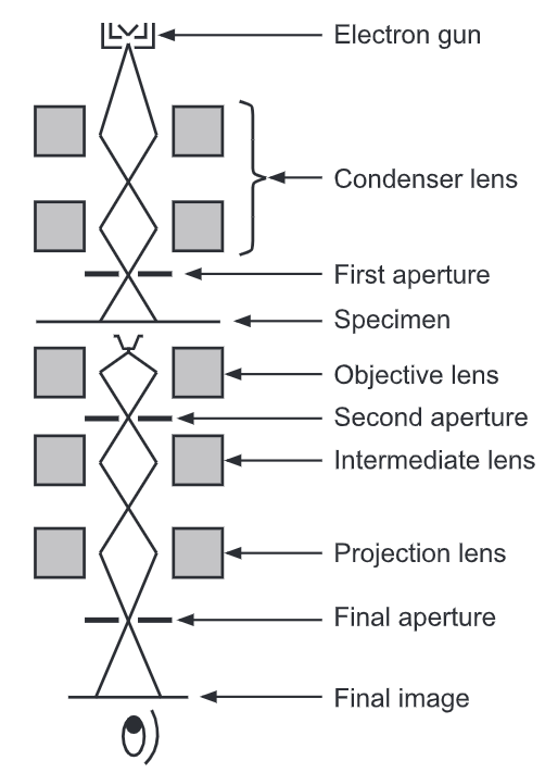
en.wikipedia.org › wiki › History_of_cell_membraneHistory of cell membrane theory - Wikipedia A more direct investigation of the membrane was made possible through the use of electron microscopy in the late 1950s. After staining with heavy metal labels, Sjöstrand et al. noted two thin dark bands separated by a light region, which they incorrectly interpreted as a single molecular layer of protein. A more accurate interpretation was ... transmission electron microscope | instrument | Britannica transmission electron microscope (tem), type of electron microscope that has three essential systems: (1) an electron gun, which produces the electron beam, and the condenser system, which focuses the beam onto the object, (2) the image-producing system, consisting of the objective lens, movable specimen stage, and intermediate and projector … The Transmission Electron Microscope | CCBER - UC Santa Barbara What is a Transmission Electron Microscope? Transmission electron microscopes (TEM) are microscopes that use a particle beam of electrons to visualize specimens and generate a highly-magnified image. TEMs can magnify objects up to 2 million times. In order to get a better idea of just how small that is, think of how small a cell is. Platelet Transmission Electron Microscopy and Flow Cytometry Platelet transmission electron microscopy (or PTEM in short) is the gold standard for assessing platelet ultra-structures such as dense and alpha granules. There are 3 main tests: Platelet whole mount TEM is to quantify dense granules. Platelet thin section TEM is the method to visualize ultrastructures such as alpha granules and inclusions.
Transmission Electron Microscope (TEM) - Uses, Advantages and Disadvantages A Transmission Electron Microscope produces images via the interaction of electrons with a sample. TEMs are costly, large, cumbersome instruments that require special housing and maintenance. They are also the most powerful microscopic tool available to-date, capable of producing high-resolution, detailed images 1 nanometer in size. ...
Electron Microscope Principle, Uses, Types and Images (Labeled Diagram ... The cost of a new electron microscope ranges between $80,000 to $10,000,000 and above depending on the customizations, configurations, resolution, components, and brand value. The type of electron microscope also decides the price of the microscope because of the various uses the microscope has and also on the components used in them.
Electron Micrographs of Cell Organelles | Zoology - Biology Discussion This is the electron micrograph of Lysosome, and is characterized by following features. These are also called Suicide bags or Death bags of the cell (Fig. 13 &14): (1) They were discovered by de Duve (1954). (2) They are spherical or irregular membrane bound vesicles filled with digestive enzymes.
en.wikipedia.org › wiki › Herpes_simplex_virusHerpes simplex virus - Wikipedia Herpes simplex virus 1 and 2 (HSV-1 and HSV-2), also known by their taxonomical names Human alphaherpesvirus 1 and Human alphaherpesvirus 2, are two members of the human Herpesviridae family, a set of viruses that produce viral infections in the majority of humans.
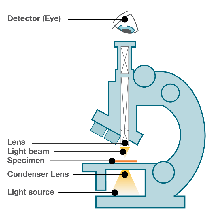
Microscope Types (with labeled diagrams) and Functions Compound Microscope. Electron Microscope. Phase-contrast Microscope. Interference Microscope. The most important feature of a microscope is that it magnifies the image of the specimen and resolves it better to help the observers see the specimen clearly to carry out their experiments. Although Zacharias and Hans Janssen are credited with the ...
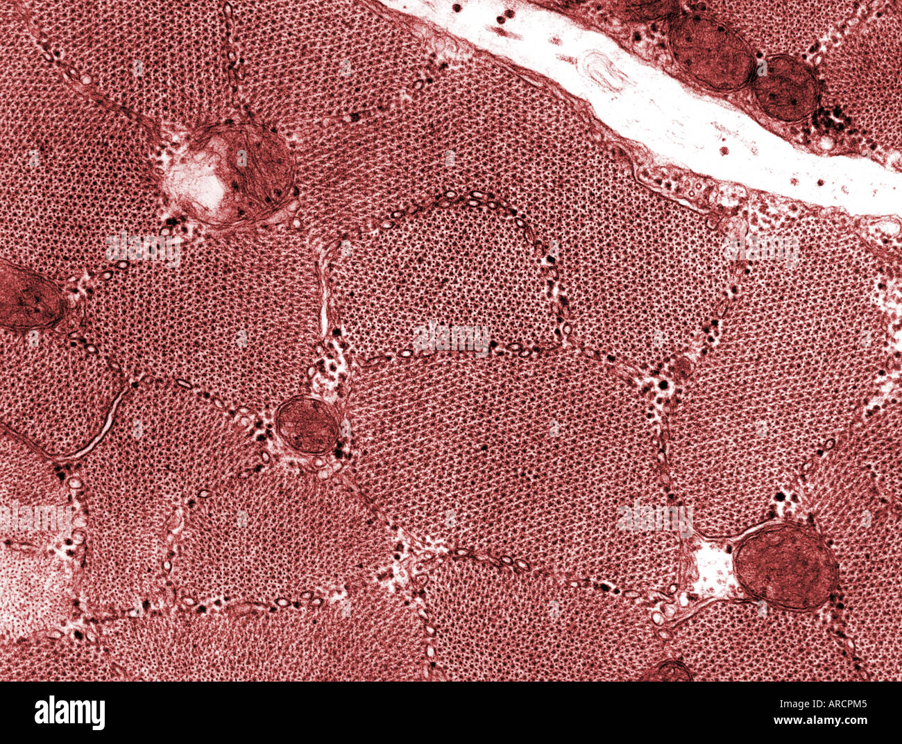
Transmission Electron Micrograph- Sarcomere Structure (Partially ... Transmission Electron Micrograph- Sarcomere Structure (Partially Contracted Muscle) Diagram | Quizlet Transmission Electron Micrograph- Sarcomere Structure (Partially Contracted Muscle) 5.0 (1 review) + − Learn Test Match Created by giordanna99 Terms in this set (6) Z disc ... M line ... H zone ... I band ... A band ... A band ... giordanna99
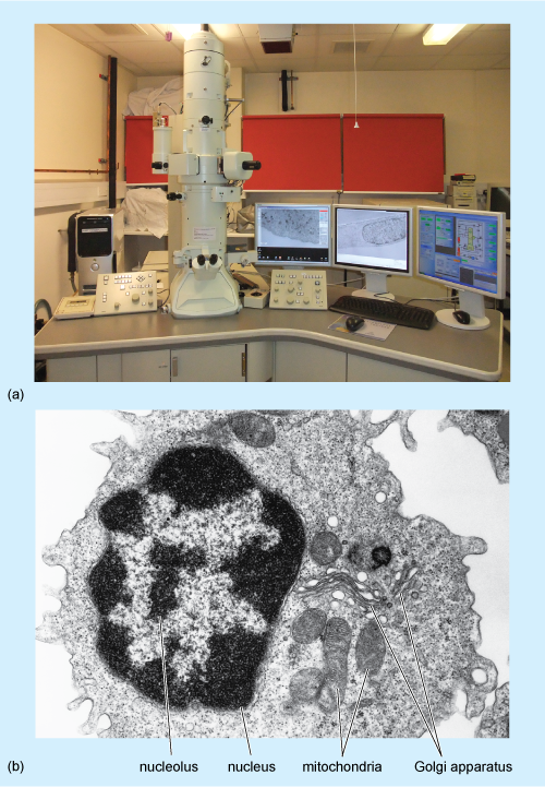
1.2 Skill: Interpretation of electron micrographs - YouTube Dec 25, 2015 ... Interpretation of electron micrographs to identify organelles and deduce the functions of specialized cells. Electron micrographs used with ...
Transmission Electron Microscopy | Materials Science | NREL Selected area transmission electron diffraction patterns can be obtained from localized regions of the sample by inserting a selected area diffraction aperture into the image plane of the objective lens.
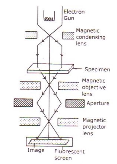
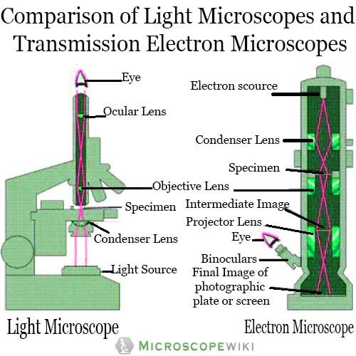
Transmission Electron Microscope (With Diagram) - Biology Discussion Finally, the electrons are focused by an electromagnetic projector lens (instead of an ocular lens as in a light microscope) on a screen or photographic plate. The final image in a TEM is known as transmission electron micrograph. The salts of some heavy metals, e.g., lead; osmium, tungsten and uranium are often used for staining.
Label the Transmission Electron Micrograph of the Nucleus Label the Transmission Electron Micrograph of the Nucleus ... Main Menu
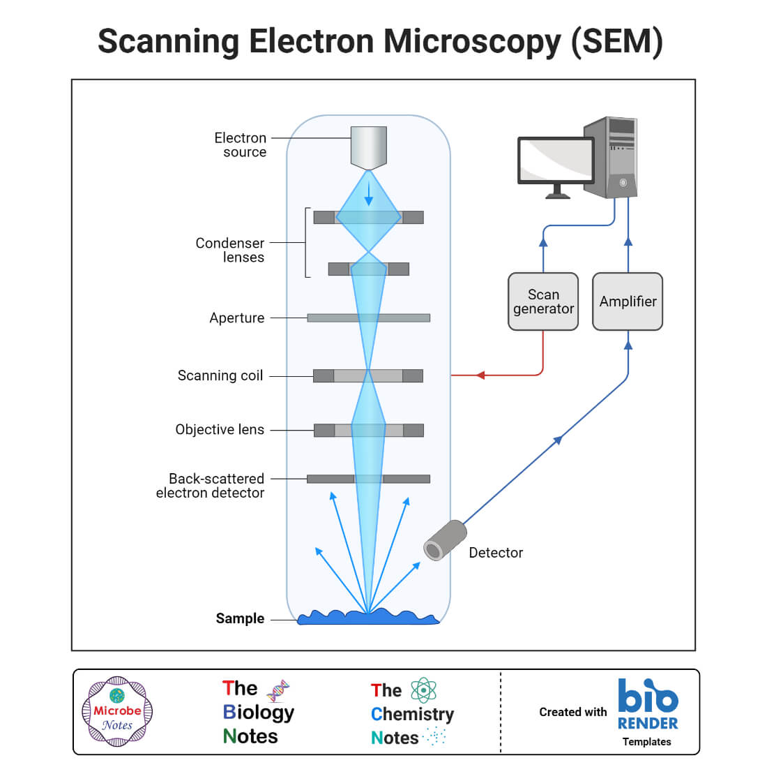
Electron Microscope- Definition, Principle, Types, Uses, Labeled Diagram There are two types of electron microscopes, with different operating styles: 1. Transmission Electron Microscope (TEM) The transmission electron microscope is used to view thin specimens through which electrons can pass generating a projection image. The TEM is analogous in many ways to the conventional (compound) light microscope.
› media › subtopicImage Library | CDC Online Newsroom | CDC This electron microscopic (EM) image depicted a mpox virion, obtained from a clinical sample associated with the 2003 prairie dog outbreak. It was a thin section image from of a human skin sample. On the left were mature, oval-shaped virus particles, and on the right were the crescents, and spherical particles of immature virions.
en.wikipedia.org › wiki › ChloroplastChloroplast - Wikipedia A chloroplast (/ ˈ k l ɔːr ə ˌ p l æ s t,-p l ɑː s t /) is a type of membrane-bound organelle known as a plastid that conducts photosynthesis mostly in plant and algal cells.The photosynthetic pigment chlorophyll captures the energy from sunlight, converts it, and stores it in the energy-storage molecules ATP and NADPH while freeing oxygen from water in the cells.
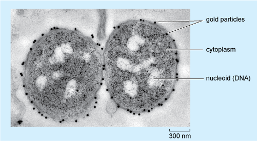
Transmission electron micrograph (TEM) identifying immunogold labeled ... Transmission electron micrograph (TEM) identifying immunogold labeled ESR1 and caveolin-1 proteins in the uterine artery endothelial cells derived from the pregnant state (P-UAEC).
› topics › agricultural-andTransmission Electron Microscopy - an overview ... 3.325.5.6 Transmission Electron Microscopy. TEM provides high-resolution imaging and is used for studying small areas or even single mineral platelets selectively. The TEM can be operated at different electron energies (often 100 keV for conventional TEM and 1 MeV for high-resolution imaging). The contrast of the TEM images is dependent on ...
Transmission Electron Micrograph of transfected HL-1 cells labeled for ... Download scientific diagram | Transmission Electron Micrograph of transfected HL-1 cells labeled for TMEM43 with immunogold. A and B. Single immunogold labeling experiments used 15 nm gold ...
en.wikipedia.org › wiki › ToxoplasmosisToxoplasmosis - Wikipedia In addition to cats, birds and mammals including human beings are also intermediate hosts of the parasite and are involved in the transmission process. However the pathogenicity varies with the age and species involved in infection and the mode of transmission of T. gondii. Toxoplasmosis may also be transmitted through solid organ transplants.
Transmission Electron Microscope - SlideShare Medium-voltage instruments work at 200-500 kV to provide a better transmission and resolution, and in high voltage electron microscopy (HVEM) the acceleration voltage is in the range 500 kV to 3 MV. Acceleration voltage determines the velocity, wavelength and hence the resolution (ability to distinguish the neighbouring microstructural features ...
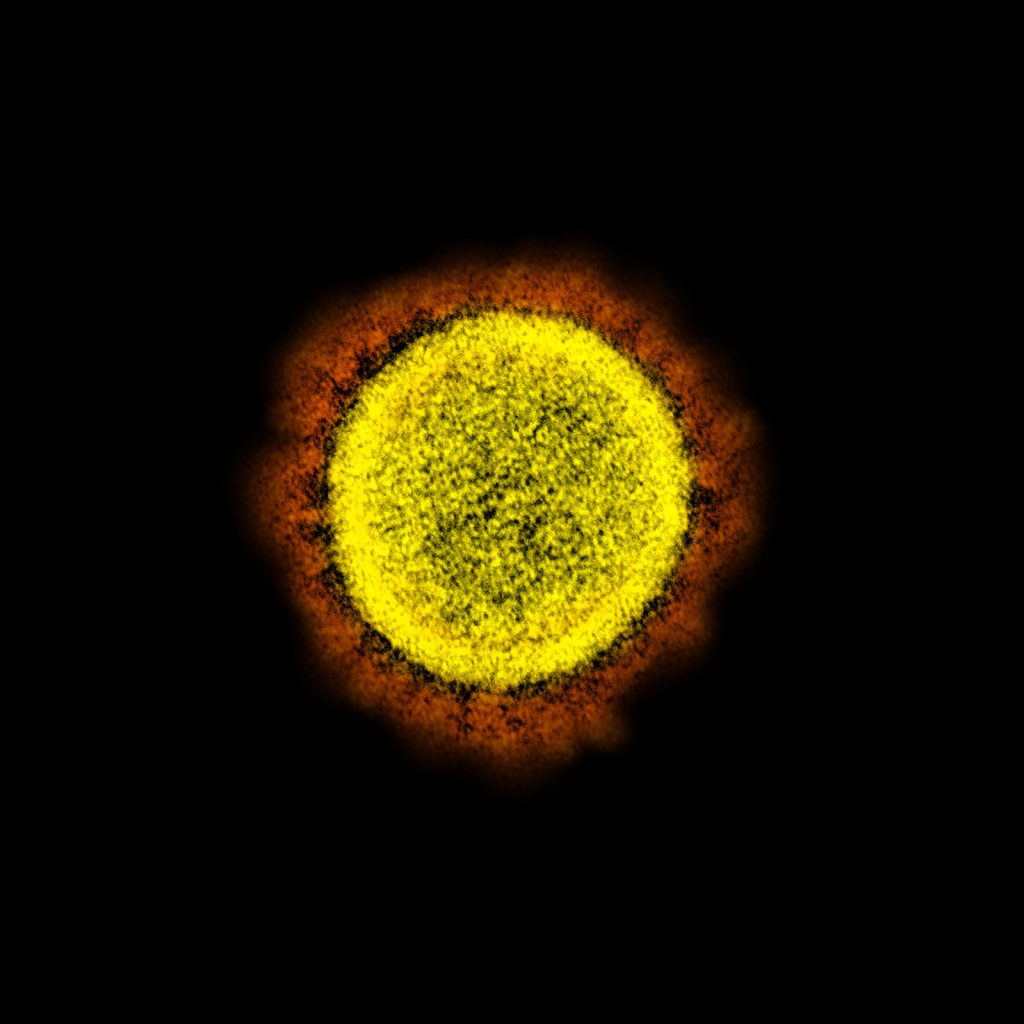
Transmission Electron Microscope (TEM)- Definition, Principle, Images Parts of a microscope with functions and labeled diagram Transmission Electron Microscope (TEM) Images Transmission electron micrograph of SARS-CoV-2 virus particles, isolated from a patient. Image captured and color-enhanced at the NIAID Integrated Research Facility (IRF) in Fort Detrick, Maryland. Credit: NIAID.
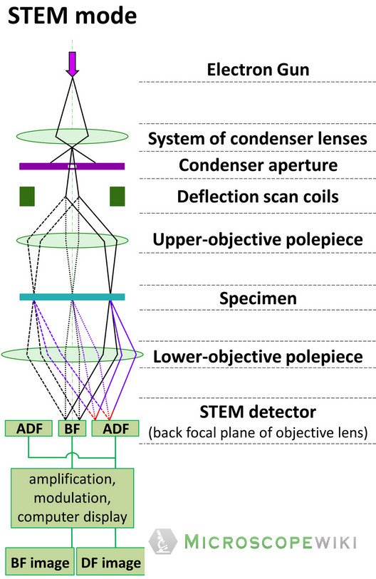
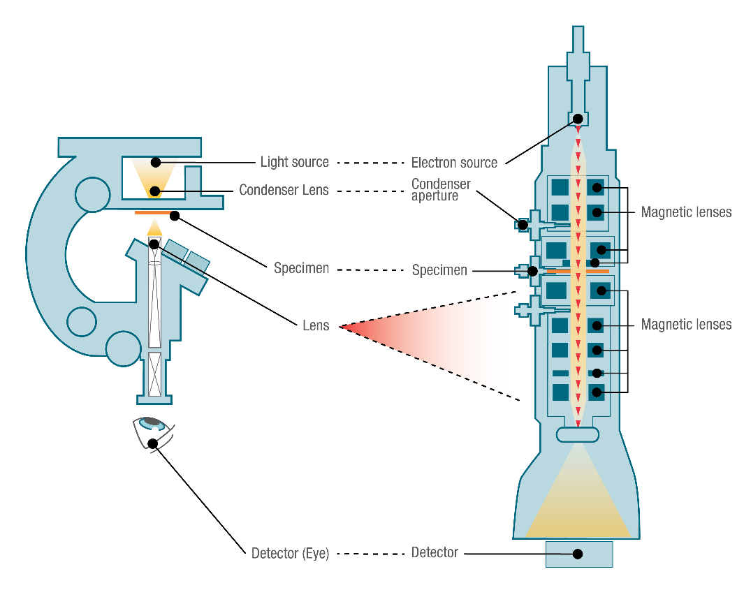
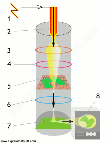
Electron Microscope-Definition, Principle, Types, Uses, Labeled Diagram The electron microscope is placed vertically and has the shape of a tall vacuum column. It consists of the following elements: 1. Electron gun. A heated tungsten filament that produces electrons makes up the electron cannon. 2. Electromagnetic lenses. The condenser lens directs the electron beam to the specimen.
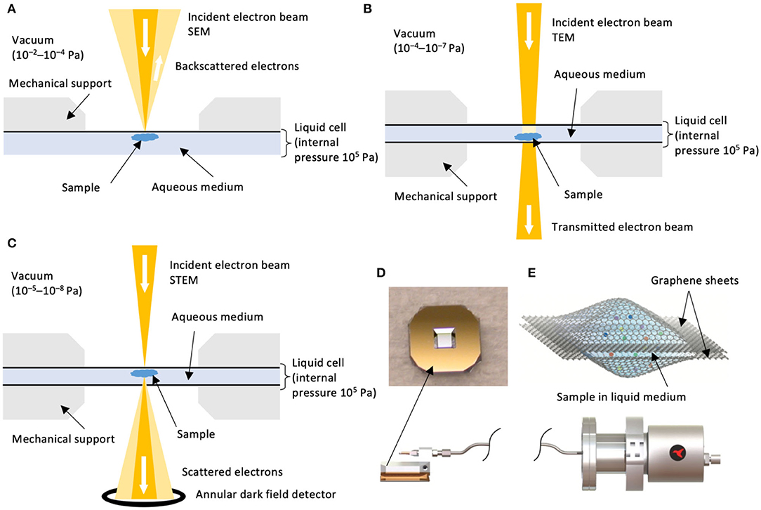
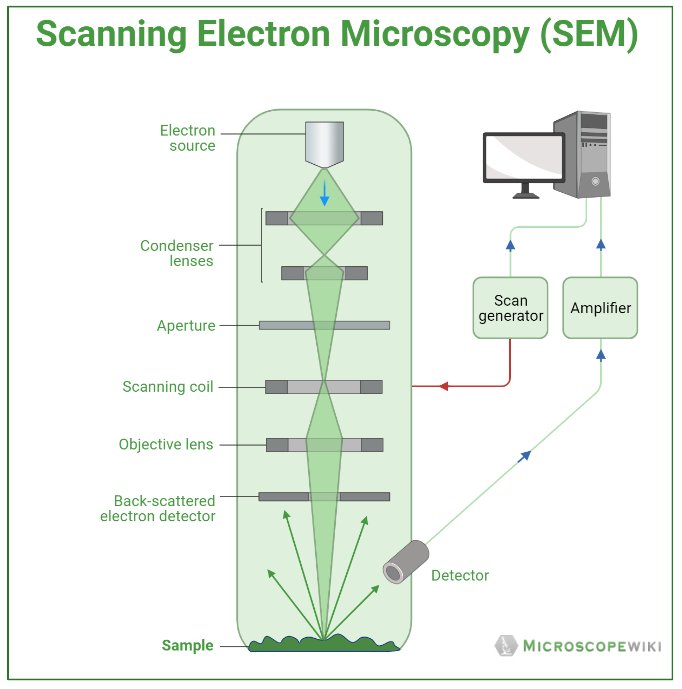
What Is an Electron Microscope (EM) and How Does It Work? - VHA ... Today there are two major types of electron microscopes used in clinical and biomedical research settings: the transmission electron microscope (TEM) and the scanning electron microscope (SEM); sometimes the TEM and SEM are combined in one instrument, the scanning transmission electron microscope (STEM):
Label This Transmission Electron Micrograph Of A Relaxed ... - Blogger Label this transmission electron micrograph of relaxed sarcomeres by clicking and dragging the labels to the correct location . Label the following image using the terms provided. Note how the sarcomeres are extended to only approximately 120 % . IMG_2132 - FIGURES Label this transmission electron from
Labeling the Cell Flashcards | Quizlet Label the transmission electron micrograph of the nucleus. membrane bound organelles golgi apparatus, mitochondrion, lysosome, peroxisome, rough endoplasmic reticulum nonmembrane bound organelles ribosomes, centrosome, proteasomes cytoskeleton includes microfilaments, intermediate filaments, microtubules Identify the highlighted structures
Transmission Electron Microscopy as a Powerful Tool to Investigate ... Despite the high resolution (3–20 nm), imaging is restricted to the surface of the sample. ... Transmission electron microscopy (TEM) provides images obtained by ...
8.2: Transmission Electron Microscopy - Chemistry LibreTexts Transmission electron microscopy (TEM) is a form of microscopy which in which a beam of electrons transmits through an extremely thin specimen, and then interacts with the specimen when passing through it. The formation of images in a TEM can be explained by an optical electron beam diagram in Figure 8.2. 1.

















Komentar
Posting Komentar